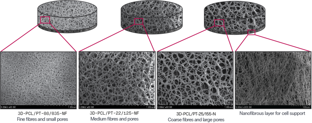
3D CELL CULTURE SCAFFOLDS

growth and interactions in all three dimensions

Product concept

3D cell culture scaffolds designed as two-layer constructs:

  • The top layer features a unique matrix of fibres and pores fabricated by the proprietary 3D fibre printing method, and serves as a matrix hosting cells. 
  • The bottom layer features a dense network of nanofibres forming a cell impenetratable membrane preventing cell migration to the well floor. 

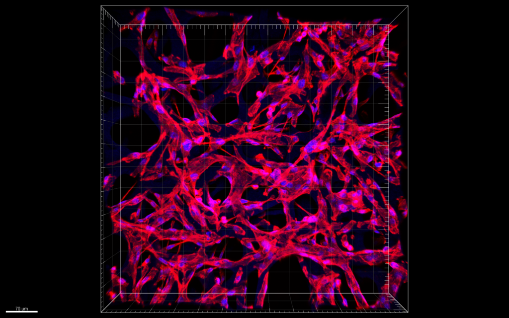
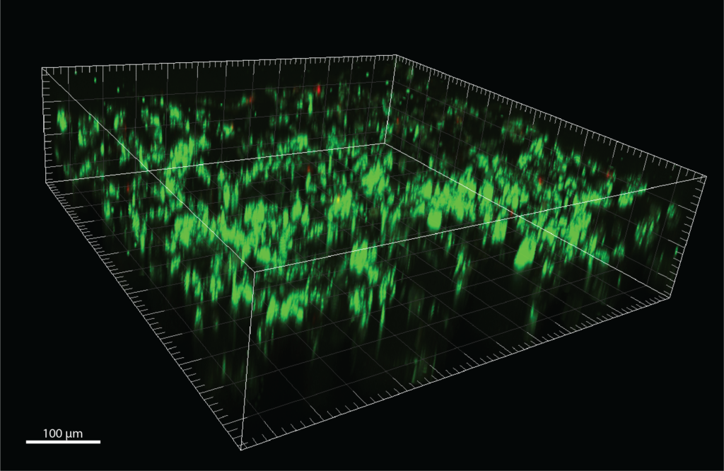
Such design provides a favourable environment for cell attachment and proliferation. The round fibre and pore morphology follows a random structure of natural extracellular matrix (as compared to straight/aligned fibre and filament structures). Large pores allow for an efficient cell distribution throughout the entire scaffold height. 

Specifications:
Construction: 3D, 2-layer (nanofibre bottom)
Basis polymer: PCL, plasma-treated

SKU: 3D-PCL/PT-08/035-NF
Fibre diameter (mean) 8 μm
Pore diameter (mean) 35 μm

SKU: 3D-PCL/PT-22/125-NF
Fibre diameter (mean) 22 μm
Pore diameter (mean) 125 μm

SKU: 3D-PCL/PT-25/155-NF
Fibre diameter (mean) 25 μm
Pore diameter (mean) 155 μm

Features
• Tunable morphology
• Cut-to-size
• Easy imaging
• UV-sterilized and ready to use
• Compatible with most current 2D assays

Applications
• Drug screening
• Environmental cytotoxicity
• Tissue engineering
• Organ-on-chip models

Modifications
• Wetting properties
• Cold plasma surface activation
• Attachment of functional molecules (growth factors)

Confocal microscopy images of MDA-MB-231 cells in scaffold 


Selectable options

  • Scaffold diameter 21, 15, 5 mm (fits 12, 24, 96 well plates, possible to use w or w/o support holders)
  • Scaffold thickness 200 ± 20 μm
  • Fibre diameter 10 – 200 μm
  • Pore diameter 15 – 500 μm
  • Morphology round interconnected pores
  • Water contact angle 40° – 70°

Method work flow

Asset 3
  1. Unpack test-plates in a laminar flow box and inspect visually.
  2. Fill wells with 70 % ethanol for 20 min and then aspirate. Wash with sterile PBS (three-times).
  3. Pre-wet wells with culture media: add culture media, incubate them for at least 30 min and aspirate the medium.
  4. Prepare cell suspension in required density and pipette the appropriate volume of cell suspension (refer to 3D cell seeding protocol).
  5. Carefully place cell suspension droplet onto the center of the wells surface.
  6. Allow droplet to distribute through the entire scaffold.
  7. Refill the wells with culture media and change the media every 3 – 4 days.
  8. Perform selected analytical procedures

Common protocols for 2D cell cultures can also be adapted when working with 3D cell cultures.
Scaffolds are compatible with both assays.

Recent application: https://doi.org/10.1016/j.bej.2022.108531

Ciuzas D., Krugly, E., Petrikaite, V. Fibrous 3D printed poly(ɛ)caprolactone tissue engineering scaffold for in vitro cell models. Biochemical Engineering Journal, Volume 185, July 2022, 108531